
Scientists have introduced a microscopy technique that reveals a hidden layer of chemistry involving molecules that normally evade detection.
A team at the University of Tokyo has created a microscopy platform that can detect a previously unseen layer of biomolecular chemistry influenced by weak magnetic fields.
The project was led by Project Researcher Noboru Ikeya and Professor Jonathan R. Woodward at the Graduate School of Arts and Sciences. Their approach tackles a long-standing challenge in life science measurements. Many key intermediates in spin-dependent reactions are “dark” molecules, meaning they do not emit light and cannot be captured with standard fluorescence imaging.
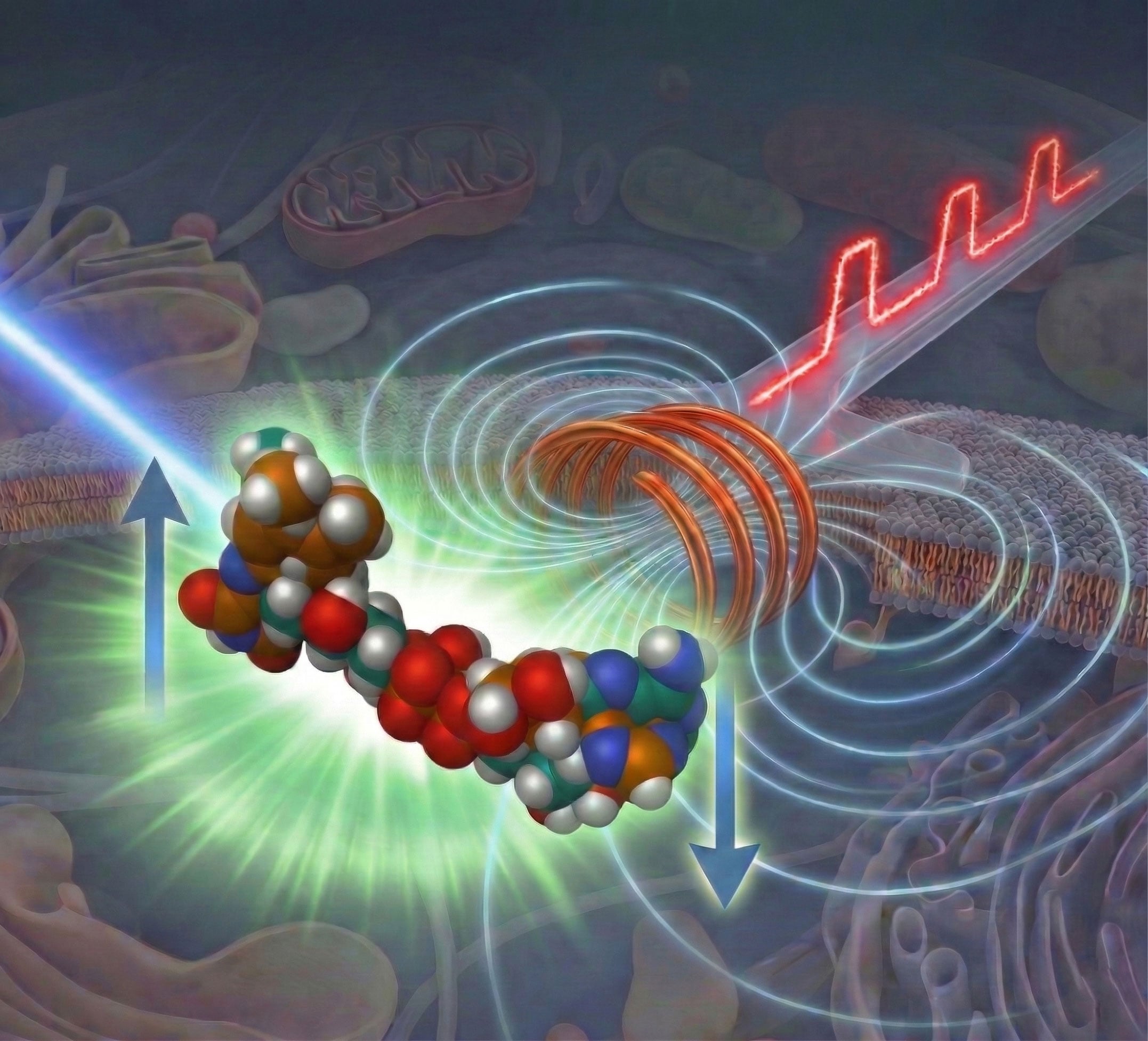
To address this limitation, the researchers combined two carefully timed light pulses with a synchronized nanosecond-scale magnetic pulse. This method, known as pump-field-probe fluorescence microscopy, tracks how signals change as the magnetic field switches at different moments in time. By comparing these changes, the system isolates the spin-dependent portion of the reaction and shows when magnetically sensitive intermediates form and vanish.
Validation in Model Systems
The team tested the technique using flavin-based model systems commonly used in studies of biologically relevant photochemistry. The results showed that the platform can measure reaction lifetimes and magnetic responses with high sensitivity, even at low concentrations similar to those found in cells.
It also detected extremely small signal variations under practical conditions that minimize sample damage, using a single experiment per frame. This capability marks an important step toward applying the method in live-cell studies.
More broadly, the work connects fluorescence microscopy with spin chemistry in a new way. It allows scientists to directly examine molecular processes that were previously inferred indirectly, offering clearer insight into how weak magnetic fields may affect biological systems. The researchers believe this approach could advance quantum biology and support the development of noninvasive diagnostic methods based on spin-sensitive molecular behavior.
Looking ahead, the team plans to apply the platform to more complex biological environments and improve analysis methods for separating overlapping reaction pathways. By making short-lived, non-emissive intermediates accessible to experiments, the technique expands what can be measured in biological photochemistry and provides a practical path for studying magnetic effects at the molecular scale.
Reference: “A Fluorescence Microscopy Platform for Time-Resolved Studies of Spin-Correlated Radical Pairs in Biological Systems” by Noboru Ikeya and Jonathan R. Woodward, 26 March 2026, Journal of the American Chemical Society.
DOI: 10.1021/jacs.5c21177
Never miss a breakthrough: Join the SciTechDaily newsletter.
Follow us on Google and Google News.